What if you had an eye on your butt?

Published by

on

One of the cool things about amphibian embryos is that the tissues heal very quickly, which enables “cut-and-paste” experiments (such as this classic) where you can move tissues within and between embryos and everything heals nicely. That allows us to ask questions about how cells and tissues function in new contexts, and to create new configurations of organ systems to see how the living material functionally adapts to novelty.

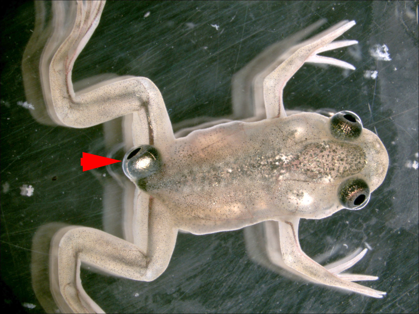
Around 2012, Douglas Blackiston, who was then a post-doc in my lab, and I began to study the behavioral and morphological consequences of eye transplants. This is different from our bioelectrically-induced eyes; what happens here is that some cells, which are normally fated to become eyes, are placed in another location on an early frog embryo and they make an eye.

Martha Constantine-Paton had done classic experiments grafting ectopic eyes, but no information was available as to whether those animals could see through those eyes. Doug is a very talented microsurgeon and developmental biologist, and was able to perfect this technique, leading to the production of tadpoles which had eyes on their tails, but not in their heads. This enabled us to ask many questions about the functional properties of this process in the context of the whole organism.

We learned many things from these experiments. First, we asked whether they could see with these etcopic eyes. How do we know what they can see? We don’t have a way of knowing what it’s like to be this animal, but we can gather evidence by observing their functional capabilities in behavioral assays that require vision. So, we built a machine that automated and quantified their ability to learn in visual tasks (avoid or follow a specific color light in the dish):

This was a huge ordeal, because many engineering challenges had to be solved (including machine vision problems, materials properties, real-time control on the millisecond timescale, etc.). The device we built was (and possibly still is) the first automated system that not only tracks animal position but automates immediate feedback to them so they can be trained. We used the same system to probe the regeneration of memories in planaria, and I think bigger versions of this robot platform could someday be used by the pharma industry in drug discovery efforts with tadpoles or zebrafish embryos to screen for nootropic drugs (and other cognitive modulators that cannot be found by screens in single cells). We discovered that the animals could see quite well, as they performed successfully in behavioral trials that required them to discriminate colors and move accordingly.

Then we asked – what do these eyes connect to?! Turns out, they don’t connect to the brain, like normal eyes do. The optic nerve that emerges from these ectopic transplanted eyes connects to the spinal cord, or sometimes the gut or sometimes to nothing obvious at all, and they can still see.

The remarkable thing is that apparently, it doesn’t take many generations of evolutionary adaptation to accommodate this change – in just 1 generation, the brain and body integrate to a new sensory-motor architecture, and the behavioral repertoire adjusts. This example of plasticity (and many others) is discussed in depth here. Exactly what it sees, and how the brain processes information arriving via the spinal cord instead of its usual route to the optic tectum, we don’t know, but it’s important for the fascinating field of sensory substitution and for understanding somatic-cognitive plasticity in general. What kind of sensors can be added to your body, with what connectors, and what sort of cognition will it enable?

We also studied the mechanisms guiding the emerging optic nerve. Turns out, like many of our other cases such as cancer cells, they are guided by the bioelectrical profile of their environment. By depolarizing the surrounding tissues, we could cause a massive increase in the amount of optic nerve. This has obvious potential relevance to regenerative therapies of the visual system. It turns out that it’s mediated by the voltage-induced communication between nerves and their environment carried by the neurotransmitter serotonin. Serotonin has many roles, including in determining body left-right asymmetry long before neurons even appear. And we found a drug – the migraine medication zolmitriptan – that can reliably induce a massive overgrowth of optic nerves. Remarkably, what it does not do is alter the growth of native nerve, even when the whole embryo is bathed in it. It’s as if only the ectopic nerves, which can somehow tell they’re out of place, are paying attention to this stimulus while those who are settled in their normal home are ignoring this information.

And the final piece of the story. One of the essential steps of tadpole development (into a frog) is to lose their tail: the tail cells are driven by hormonal signals to self-destruct. So now, the key question was: what would happen to the ectopic eye on a tail, as the tail around it degrades?

We had a poll in the lab, for guesses as to whether it would disappear or not. What would you guess? The actual answer? Here it is:

The eye ignores the programmed carnage around it – hormones and all – because apparently it knows that those death signals are not meant for it. It thrives, grows, and moves anteriorly as the tail shrivels, eventually finding its proper place on the mature frog’s posterior.

Have I received emails afterward, from people asking me to give them an eye on their butt? Yes, yes I have.

12 responses to “What if you had an eye on your butt?”

  1. chris m Avatar
    chris m

    can that be inherited?

    1. Mike Levin Avatar
      Mike Levin

      In frogs, we don’t know (but it’s not super likely). In planaria, yes, we’ve made 2-headed worms and that’s inherited in their normal mode of reproduction (fission and regeneration).

  2. Ben Moskowitz Avatar
    Ben Moskowitz

    Awesome work! This is such a bizarre, fascinating example of plasticity, especially that the eye is retained in the frog! This reminds me of how when humans lose a sense some of the other senses get enhanced to compensate. Or even a story I’ve read about a person who was bling a taught himself to see through echolocation. Organisms can often display remarkable adaptation to drastic changes in their sensory landscape. I’m excited to see these ideas being experimented with to see if one day we can engineer such adaptations!

  3. Harry Buck Avatar

    Given that you can induce optic nerve development, have you considered answering your questions about “other sensors” by, for instance (surgically) replacing the ectopic eye with a different sensor (e.g. infrared, RF, etc) and testing for its functionality?

    1. Mike Levin Avatar
      Mike Levin

      Yep, we’re doing all kinds of stuff like that, giving biological organisms (and cells) new affordances – novel sensors, effectors, etc. Bernd Fritzsch (https://pubmed.ncbi.nlm.nih.gov/?term=Fritzsch+B&cauthor_id=35134251) has done some stuff on transplants eye/ear in Xenopus.

    2. Mina Basily Avatar
      Mina Basily

      Remarkable and outstanding work, Dr. Levin,

      I have a question about the case when there was no obvious neural linkage from the eye to anywhere in the body and the organism was still able to see.

      Do you think somehow the non-excitable cells that surround the ectopic eye in this model have gained an extra feature during development that makes them able to transport the information from the ectopic eye to the brain to compensate the absence of the optic nerve?
      I think If this is somehow what happened, that would be an outstanding example of cellular plasticity.

  4. Brian P Avatar
    Brian P

    Love your work and insights!

    What are the implications of this bioelectric induced morphological capability with respect to human eye conditions such as myopia (impacts 1.45 billion people globally)?

    Could there be an ion channel pathology involved in myopia? From the initial research I did, it looks like there is a connection:

    Biochemistry and Molecular Biology | June 2023
    The Mechanosensitive Piezo1 Channel Mediates Mechanochemical Transmission in Myopic Eyes
    Weiqi Zhong; Changjun Lan; Zhiming Gu; Qingqing Tan; Xiaoling Xiang; Hong Zhou; Xuan Liao

    https://iovs.arvojournals.org/article.aspx?articleid=2785666

    Disrupted potassium ion homeostasis in ciliary muscle in negative lens-induced myopia in Guinea pigs
    May 2020
    Archives of Biochemistry and Biophysics 688(6):108403
    Shanshan Wu a, Dadong Guo b, Huixia Wei a, Xuewei Yin a, Liwei Zhang a, Bin Guo a, Furu Xu a, Yixian Hao a, Wenjun Jiang b, Hongsheng Bi

    https://linkinghub.elsevier.com/retrieve/pii/S0003986120304124

    The role of voltage-gated ion channels in visual function and disease in mammalian photoreceptors.
    Rabab Rashwan1, Rabab Rashwan2, David M. Hunt2, David M. Hunt3 +2 more•Institutions (3)
    12 Jul 2021-Pflügers Archiv: European Journal of Physiology (Springer Berlin Heidelberg)-Vol. 473, Iss: 9, pp 1455-1468
    Advances in biomedical study of the myopia-related signaling pathways and mechanisms
    Jing Yang a, Xinli Ouyang a, Hong Fu a, Xinyu Hou a, Yan Liu b, Yongfang Xie a, Haiqun Yu b, Guohui Wang a

    There are also some alternative therapies (red light laser therapy and electrical stimulation) that suggest myopia is modifiable:

    Effect of Repeated Low-level Red Light on Myopia Prevention Among Children in China With Premyopia
    A Randomized Clinical Trial
    Xiangui He, PhD1,2; Jingjing Wang, PhD1; Zhuoting Zhu, PhD3,4,5; et al
    JAMA Netw Open. 2023;6(4):e239612. doi:10.1001/jamanetworkopen.2023.9612

    Efficacy of Repeated Low-Level Red-Light Therapy for Slowing the Progression of Childhood Myopia: A Systematic Review and Meta-Analysis

    Jie Tang, Ya Liao, Na Yan, Shiferaw Blen Dereje, Jingjing Wang, Yunjiao Luo, Yuhao Wang, Wen Zhou, Xiaojuan Wang, Wei Wang

    Ciliary Muscle Electrostimulation to Restore Accommodation in Patients With Early Presbyopia: Preliminary Results
    Luca Gualdi, MD, Federica Gualdi, MD, Dario Rusciano, PhD, Renato Ambrósio Jr., MD, PhD, Marcella Q. Salomão, MD, Bernardo Lopes, MD, Veronica Cappello, MD, Tatiana Fintina, MD, and Massimo Gualdi, MD
    Journal of Refractive Surgery, 2017;33(9):578–583

    Electrical Stimulation as a Means for Improving Vision.
    Amer Sehic1, Shuai Guo2, Kin-Sang Cho3, Rima Maria Corraya2 +2 more•Institutions (3)
    31 Oct 2016-American Journal of Pathology (Elsevier)-Vol. 186, Iss: 11, pp 2783-2797

    I’m curious to hear your thoughts on the possibilities of targeting ion channels with electroceuticals to reverse myopia.

  5. Dr. Niko Papazoglou Avatar

    As I often emphasize to my patients, we don’t see with our eyes but with our brains. While our eyes receive light reflected or emitted from objects, they simply convert this light into neural signals. The true perception of these signals as images only occurs after intricate processing by the brain.

    In the case of humans, having two eyes means that the images received from each eye, due to their slightly different angles of reception, allow our brains to construct a stereoscopic image.

    However, deviations from this norm, such as in cases of strabismus, which your “eye on the butt” scenario is equivalent to, result in the eyes looking in totally different directions. Consequently, the brain cannot merge the images into a cohesive 3D image. As a result, the brain permanently attenuates the weaker and less relevant image, even if the eye is physically functioning. This condition is known medically as amblyopia or lazy eye to the public.

    One of my points is that simply copying and pasting eyes onto a living organism isn’t sufficient for developing a functional visual system. Evolution must be considered, particularly in higher-level organisms, where eyes are more complex than simple reactive photoreceptors.

    While this work is intriguing, it remains light years away from being truly meaningful in medical practice.

    1. Mike Levin Avatar
      Mike Levin

      This is an interesting point, but if people can learn to see (even if a partial degree) with tongue and skin interfaces (Bach-y-Rita’s work and more recent “electric lollipop” work), ectopic eyes can be made to work too. I’m in no way claiming our frog work is currently ready for medical practice. Our initial reason for doing it is to learn something about evolution and behavioral plasticity. But I think there is a path for clinical sensory augmentation not only with normal eyes but with much more alien interfaces.

  6. Benjamin L Avatar
    Benjamin L

    On a conceptual level, this reminds me of the work of Vernon Smith, who won a Nobel Prize for experimental economics. His experiments found that markets were able to achieve efficient outcomes despite deviating from the precise starting conditions that economists had presumed were necessary.

    Here’s a very short article on Vernon Smith’s work: https://www.imf.org/external/pubs/ft/fandd/2003/03/clif.htm

  7. Nina Alexieva Avatar
    Nina Alexieva

    Could that mean that I can see with an eye if the optical nerve is damaged? If it (the eye) can attach on other than the brain – could it also repair the optic nerve?

    1. Mike Levin Avatar
      Mike Levin

      We don’t know how all of this will play out with mammals (especially using the human-approved drug we found to increase optic nerve growth in tadpoles) so maybe, but we’re not sure yet.

Leave a Reply to Mike Levin Cancel reply

Your email address will not be published. Required fields are marked *